VESTIBULAR DISEASE IN ANIMALS
by R.M. Clemmons, DVM, PhD
Associate Professor of Neurology & Neurosurgery
Introduction:
All veterinary species suffer from various forms of vestibular disease.
Many of which require only recognition, while others represent significant
diagnostic challenges. Although there are a number of diseases which can
affect the vestibular system, generally we can break them down anatomically
into peripheral and central disorders. With certain exceptions, peripheral
diseases bear a better prognosis in most species than central vestibular
disease. Partially due to this concern, vestibular diseases represent a
large number of neurologic referrals. Often, it is only reassurance that
the problem will pass that is necessary. Recognition of when to intervene
is as important as when not too.
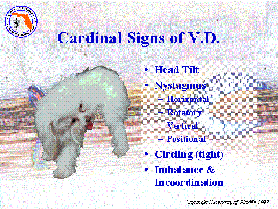 The cardinal
signs of unilateral vestibular disease are head tilt, nystagmus (spontaneous
abnormal eye movements), circling (toward the lesion in "tight" circles),
and incoordination. This is because the vestibular system is an important
part of the CNS balance control system. In order for animals to know how
they are oriented in space, three neural systems must be functioning. The
vestibular system, through the stimulus-response of the hair cells in the
semicircular canals, reacts to angular acceleration and deceleration. The
visual system allows the animal to focus on the horizontal and vertical,
orienting in space. Finally, gravity is detected by pressure receptors
in the skin, orienting the animal on up and down. While the vestibular
system is very important, it requires at least 2 of these orienting systems
to function for the animal to negotiate within its environment. This can
be important with vestibular disease, since, in acute disease, the nystagmus
prevents the eyes from focusing on the horizon, effectively eliminating
spacial orientation.
The cardinal
signs of unilateral vestibular disease are head tilt, nystagmus (spontaneous
abnormal eye movements), circling (toward the lesion in "tight" circles),
and incoordination. This is because the vestibular system is an important
part of the CNS balance control system. In order for animals to know how
they are oriented in space, three neural systems must be functioning. The
vestibular system, through the stimulus-response of the hair cells in the
semicircular canals, reacts to angular acceleration and deceleration. The
visual system allows the animal to focus on the horizontal and vertical,
orienting in space. Finally, gravity is detected by pressure receptors
in the skin, orienting the animal on up and down. While the vestibular
system is very important, it requires at least 2 of these orienting systems
to function for the animal to negotiate within its environment. This can
be important with vestibular disease, since, in acute disease, the nystagmus
prevents the eyes from focusing on the horizon, effectively eliminating
spacial orientation.
 The anatomic
structures involved in the vestibular system include the hair cells
in the saccule and utricle (containing the semicircular canals), the vestibular
portion of CN VIII, the vestibular nuclei in the brainstem and the flocculonodular
lobe of the cerebellum. The vestibular nuclei send fibers forward in the
medial longitudinal fasciculus (MLF) which coordinates ocular movements,
projects fibers to the spinal cord as the vestibulospinal tract and descending
MLF, projects fibers to the cerebellum, and sends fibers to various structures
in the brainstem including the emetic center. Involvement of any of the
portions of the vestibular system will result in signs of disfunction.
Most lesions result in loss of function and, hence, are ablative in nature.
The signs develop due to the imbalance existing between the normal and
abnormal sides.
The anatomic
structures involved in the vestibular system include the hair cells
in the saccule and utricle (containing the semicircular canals), the vestibular
portion of CN VIII, the vestibular nuclei in the brainstem and the flocculonodular
lobe of the cerebellum. The vestibular nuclei send fibers forward in the
medial longitudinal fasciculus (MLF) which coordinates ocular movements,
projects fibers to the spinal cord as the vestibulospinal tract and descending
MLF, projects fibers to the cerebellum, and sends fibers to various structures
in the brainstem including the emetic center. Involvement of any of the
portions of the vestibular system will result in signs of disfunction.
Most lesions result in loss of function and, hence, are ablative in nature.
The signs develop due to the imbalance existing between the normal and
abnormal sides.
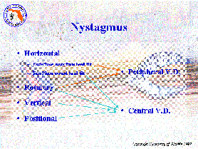
The nystagmus seen in vestibular disease can 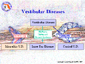 be
helpful in localizing the disease process. While horizontal and rotatory
nystagmus can be seen with disease anywhere within the vestibular system,
vertical and positional nystagmus are almost exclusively seen with central
vestibular diseases. Moreover, horizontal nystagmus from peripheral vestibular
disease oscillates with the fast-phase away from the direction of the head
tilt. With central vestibular disease (particularly of the cerebellum),
however, the fast-phase is toward the lesion. So although horizontal and
rotatory nystagmus are not specific for peripheral disease, they are compatible
with it. Vertical and positional nystagmus suggest the lesion is within
the CNS and indicate the need for a thorough neurologic work-up.
be
helpful in localizing the disease process. While horizontal and rotatory
nystagmus can be seen with disease anywhere within the vestibular system,
vertical and positional nystagmus are almost exclusively seen with central
vestibular diseases. Moreover, horizontal nystagmus from peripheral vestibular
disease oscillates with the fast-phase away from the direction of the head
tilt. With central vestibular disease (particularly of the cerebellum),
however, the fast-phase is toward the lesion. So although horizontal and
rotatory nystagmus are not specific for peripheral disease, they are compatible
with it. Vertical and positional nystagmus suggest the lesion is within
the CNS and indicate the need for a thorough neurologic work-up.
Vestibular diseases can be classified into three major disease
processes: idiopathic vestibular disease, inner ear disease, or central
vestibular disease. The former 2 represent common forms of peripheral vestibular
disease which need to be separated from each other and from central vestibular
disease.
Idiopathic Vestibular Disease:
All cranial nerves have the potential to develop specific syndromes
which are clinically classified as idiopathic disorders. This is probably
due to the fact that each cranial nerve represents a unique developmental
anatomy from their respective brachial arches. This also gives them an
unique antigenic signal allowing very specific immune attack upon them.
Idiopathic vestibular disease represents one of these cranial nerve syndromes.
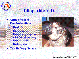 Clinically,
idiopathic vestibular disease presents as an acute onset of vestibular
signs with severe imbalance, due to its sudden onset and the severe nystagmus
which is associated with the onset of the disorder. Since the eyes are
unable to fix on the horizon and the vestibular mechanism is defective,
there is severe vertigo. This often results in the rolling and rolling
described by the owners. This can be mistaken for a seizure, which it is
not. During the early phases of idiopathic vestibular disease, the patient
often experiences nausea to the point of frequent vomiting and inappetence.
The head tilt will be toward the side of dysfunction and the nystagmus
will be horizontal or rotatory with the fast-phase away from the head tilt.
If supported, there are no other neurologic deficits and proprioception
is normal.
Clinically,
idiopathic vestibular disease presents as an acute onset of vestibular
signs with severe imbalance, due to its sudden onset and the severe nystagmus
which is associated with the onset of the disorder. Since the eyes are
unable to fix on the horizon and the vestibular mechanism is defective,
there is severe vertigo. This often results in the rolling and rolling
described by the owners. This can be mistaken for a seizure, which it is
not. During the early phases of idiopathic vestibular disease, the patient
often experiences nausea to the point of frequent vomiting and inappetence.
The head tilt will be toward the side of dysfunction and the nystagmus
will be horizontal or rotatory with the fast-phase away from the head tilt.
If supported, there are no other neurologic deficits and proprioception
is normal.
The diagnosis of idiopathic vestibular disease is tentatively made by
the presence of acute clinical signs in the absence of other physical findings.
The minimum data base include physical examination, otoscopic examination
and neurologic examination. The lack of findings (other than the peripheral
vestibular signs) supports the 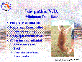 diagnosis.
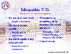
The signs of idiopathic vestibular disease are regressive, meaning that
they disappear without treatment over time. As such, the fact that the
disease is self-limiting is how the final diagnosis is achieved. The nystagmus
will usually improve or disappear all together within 3-5 days of the onset.
The patient will, then, improve in their imbalance and be more able to
function normally. This improvement will continue until minimal deficits
will remain. It is possible that there will be a residual head tilt. If
the head tilt persists beyond 6 months following the onset of signs, it
is likely to be permanent.
diagnosis.
The signs of idiopathic vestibular disease are regressive, meaning that
they disappear without treatment over time. As such, the fact that the
disease is self-limiting is how the final diagnosis is achieved. The nystagmus
will usually improve or disappear all together within 3-5 days of the onset.
The patient will, then, improve in their imbalance and be more able to
function normally. This improvement will continue until minimal deficits
will remain. It is possible that there will be a residual head tilt. If
the head tilt persists beyond 6 months following the onset of signs, it
is likely to be permanent.
 There is no
treatment which will hasten the recovery from idiopathic vestibular disease.
Corticosteroids probably do not offer an effective treatment. On the other
hand, since idiopathic vestibular disease may represent an immune disease,
anti-oxidant steroids (such as Solu Medral) may decrease severe symptoms.
During the early phases, anti-vertigo drugs might make the patient more
comfortable. Generally, I use diphenhydramine at 2-4 mg/kg every 8 hours
as needed. Diphenhydramine is a centrally active anticholinergic, antihistamine
which helps reduce vertigo and nausea. Assuming that the regressive course
becomes evident, then I monitor the patient periodically for the signs
of continued improvement.
There is no
treatment which will hasten the recovery from idiopathic vestibular disease.
Corticosteroids probably do not offer an effective treatment. On the other
hand, since idiopathic vestibular disease may represent an immune disease,
anti-oxidant steroids (such as Solu Medral) may decrease severe symptoms.
During the early phases, anti-vertigo drugs might make the patient more
comfortable. Generally, I use diphenhydramine at 2-4 mg/kg every 8 hours
as needed. Diphenhydramine is a centrally active anticholinergic, antihistamine
which helps reduce vertigo and nausea. Assuming that the regressive course
becomes evident, then I monitor the patient periodically for the signs
of continued improvement.
Antidotal evidence suggests that idiopathic vestibular disease may represent
toxicity to eating certain strains of lizards. Owners often notice the
cat with a lizard in its mouth just prior to the onset of clinical signs.
However, experimental feeding of the suggested lizard species to cats does
not lead to the disease. It is still possible that laboratory conditions
do not mimic field conditions. On the other hand, idiopathic vestibular
disease occurs in many animals and in animal species where exposure to
lizards plays no role in the condition. It is most likely that idiopathic
vestibular disease is an immune-related condition affecting the unique
antigens presented by the vestibular nerve. It can recur and is often more
severe on recurrence.
Inner Ear Disease:
Many different problems result in inner ear disease; however, the clinical
signs caused by these diseases are similar, indicating the location of
the disease rather than the specific cause. These signs are those of peripheral
vestibular dysfunction, including head tilt, nystagmus, circling and imbalance.
On the other hand, since the diseases which cause inner ear disease are
usually slower in evolution, these signs are generally less severe than
with idiopathic vestibular disease. In addition to the vestibular signs,
there are also varying degrees of facial nerve dysfunction and often Horner's
syndrome. Anatomically, the facial nerve and the sympathetic fibers heading
to the eye pass near the 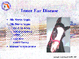 inner
ear in the osseous petrous temporal bone. Damage of these neural structure,
in addition to the damage of the vestibular nerve is a hallmark for inner
ear disease. It is possible to affect both the facial and vestibular nerves
together in the calivarium, but it is rare to see Horner's syndrome from
central nervous system disease. As such, Horner's syndrome suggests that
the disease in process is in the peripheral C8-T2 nerve roots, the vagosympathetic
trunk, the inner ear or within the orbit. When Horner's syndrome is seen
in combination with vestibular disease and facial nerve disease, the location
must be in the peripheral vestibular system in the region of the osseous
petrous temporal bone.
inner
ear in the osseous petrous temporal bone. Damage of these neural structure,
in addition to the damage of the vestibular nerve is a hallmark for inner
ear disease. It is possible to affect both the facial and vestibular nerves
together in the calivarium, but it is rare to see Horner's syndrome from
central nervous system disease. As such, Horner's syndrome suggests that
the disease in process is in the peripheral C8-T2 nerve roots, the vagosympathetic
trunk, the inner ear or within the orbit. When Horner's syndrome is seen
in combination with vestibular disease and facial nerve disease, the location
must be in the peripheral vestibular system in the region of the osseous
petrous temporal bone.
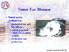 The signs of
facial nerve dysfunction include paresis or paralysis of the muscles of
facial expressions (lack of ear movement, lack of blink and lack of buccal
muscle reaction on palpation). This leads to deficiency of the vibrissa
reaction, decreased to absent menace response and diminished to absent
palpebral response. In addition, the facial nerve supplies parasympathetic
innervation to the lacrimal gland of the eye. As such, peripheral facial
nerve disease can lead to diminished tear production in the eye on the
affected side. This can be rather catastrophic in inner ear disease where
the facial nerve dysfunction results in the inability to close the eye,
while also decreasing tear production. As such, every dog with inner ear
disease should have a Schirmer's tear test run on the eyes and appropriate
treatment instituted if tear production is deficient.
The signs of
facial nerve dysfunction include paresis or paralysis of the muscles of
facial expressions (lack of ear movement, lack of blink and lack of buccal
muscle reaction on palpation). This leads to deficiency of the vibrissa
reaction, decreased to absent menace response and diminished to absent
palpebral response. In addition, the facial nerve supplies parasympathetic
innervation to the lacrimal gland of the eye. As such, peripheral facial
nerve disease can lead to diminished tear production in the eye on the
affected side. This can be rather catastrophic in inner ear disease where
the facial nerve dysfunction results in the inability to close the eye,
while also decreasing tear production. As such, every dog with inner ear
disease should have a Schirmer's tear test run on the eyes and appropriate
treatment instituted if tear production is deficient.
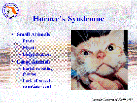 Horner's syndrome
varies among species. In small animals the ocular signs predominate, including
myosis, ptosis and enophthalmos. In horses, the signs of Horner's syndrome
are expressed primarily as excessive sweating on the affected side of the
face. In cattle, there is a lack of sweating on the muzzle of the affected
side.
Horner's syndrome
varies among species. In small animals the ocular signs predominate, including
myosis, ptosis and enophthalmos. In horses, the signs of Horner's syndrome
are expressed primarily as excessive sweating on the affected side of the
face. In cattle, there is a lack of sweating on the muzzle of the affected
side.
The most common cause of inner ear disease in all species is secondary
in inner ear infection. Most of these represent bacterial extension from
otitis media which can arise from hematogenous spread from bacteremia or
from spread up the eustachian tube to the middle ear. Luckily, these infections,
once recognized, can often be successfully treated. Other causes of inner
ear disease may not be treatable, including fungal infections and neoplasia.
Therefore, it is generally best to "treat-for-the-treatable" when dealing
with inner ear disease, using appropriate antibiotic therapy.
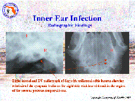 The minimum
data base for diagnosis of inner ear disease includes physical and neurologic
examination, Schirmer's tear test, otoscopic examination (with culture
of the external ear canal, if indicated), pharyngeal examination, CBC and
urinalysis. If there is evidence of cardiac murmur, then cardiac ultrasound
should be performed. Skull radiographs are then necessary to evaluate the
degree of change in the osseous structures of the inner ear. This will
be helpful in making the diagnosis and in monitoring the response to treatment.
The minimum
data base for diagnosis of inner ear disease includes physical and neurologic
examination, Schirmer's tear test, otoscopic examination (with culture
of the external ear canal, if indicated), pharyngeal examination, CBC and
urinalysis. If there is evidence of cardiac murmur, then cardiac ultrasound
should be performed. Skull radiographs are then necessary to evaluate the
degree of change in the osseous structures of the inner ear. This will
be helpful in making the diagnosis and in monitoring the response to treatment.
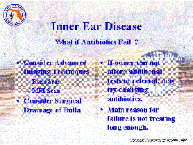 The treatment
of bacterial inner ear infection must consider the fact that the disease
represents bone infection. As such, the antibiotic must be able to penetrate
bone, develop good tissue concentrations (including the blood-ear barrier)
and, preferably, be bactericidal in action. Many veterinarians use enrofloxacin
as their antibiotic of choice. I find that enrofloxacin is great for treating
gram negative infections in the lung, but it may not reach tissue concentrations
within neural structures like the inner ear. It needs additional help to
do this. Therefore, if I do not use my treatment of choice, I will add
a sulfa drug to enrofloxacin to take advantage of the synergistic effect
of sulfa drugs. My antibiotic regime of choice is cephalosporins and sulfa
drugs (sulfadimethozine) in combination. This meets the criteria for the
ideal therapy for inner ear disease. It is excellent in treating gram positive
bacteria, which are the most common organisms infecting the inner ear.
Since this is a bone infection, the treatment must be continued for 6-8
weeks, minimum. The most common cause of treatment failure is not treating
long enough.
The treatment
of bacterial inner ear infection must consider the fact that the disease
represents bone infection. As such, the antibiotic must be able to penetrate
bone, develop good tissue concentrations (including the blood-ear barrier)
and, preferably, be bactericidal in action. Many veterinarians use enrofloxacin
as their antibiotic of choice. I find that enrofloxacin is great for treating
gram negative infections in the lung, but it may not reach tissue concentrations
within neural structures like the inner ear. It needs additional help to
do this. Therefore, if I do not use my treatment of choice, I will add
a sulfa drug to enrofloxacin to take advantage of the synergistic effect
of sulfa drugs. My antibiotic regime of choice is cephalosporins and sulfa
drugs (sulfadimethozine) in combination. This meets the criteria for the
ideal therapy for inner ear disease. It is excellent in treating gram positive
bacteria, which are the most common organisms infecting the inner ear.
Since this is a bone infection, the treatment must be continued for 6-8
weeks, minimum. The most common cause of treatment failure is not treating
long enough.
Central Vestibular Disease:
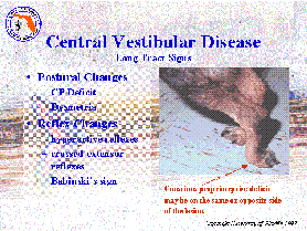 Whenever anything
else is seen other than the signs above, one must consider the likelihood
that the problem is due to central vestibular disease. Additional cranial
nerve deficits, proprioceptive deficits and motor deficits indicate brainstem
damage affecting the vestibular nuclei and sensor and motor pathways which
course through the vestibular region of the brainstem. In addition, the
nystagmus seen in central vestibular disease will often be vertical or
positional in nature, supporting the location of the disease process within
the brainstem or cerebellum. If there is whole body and head tremors, the
lesion is likely to be within the flocculonodular lobe of the cerebellum.
While diseases which affect the peripheral vestibular system are usually
good diseases; that is, diseases which regress without treatment or which
respond to appropriate antibiotic therapy, most central vestibular diseases
carry a less optimistic prognosis.
Whenever anything
else is seen other than the signs above, one must consider the likelihood
that the problem is due to central vestibular disease. Additional cranial
nerve deficits, proprioceptive deficits and motor deficits indicate brainstem
damage affecting the vestibular nuclei and sensor and motor pathways which
course through the vestibular region of the brainstem. In addition, the
nystagmus seen in central vestibular disease will often be vertical or
positional in nature, supporting the location of the disease process within
the brainstem or cerebellum. If there is whole body and head tremors, the
lesion is likely to be within the flocculonodular lobe of the cerebellum.
While diseases which affect the peripheral vestibular system are usually
good diseases; that is, diseases which regress without treatment or which
respond to appropriate antibiotic therapy, most central vestibular diseases
carry a less optimistic prognosis.
 The major causes
of central vestibular disease are inflammatory/infectious diseases or neoplasia.
Organophosphate intoxication, liver disease (with metabolic brainstem degeneration)
and thiamine deficiency can occasionally result in central vestibular disease
(depending upon the species of animal), but these causes are far less than
the inflammatory or neoplastic causes. In dogs, canine distemper virus,
granulomatous meningoencephalitis, toxoplasmosis, neosporidiosis, aspergillosis,
cryptococcosis, steroid-responsive meningoencephalitis, Lyme's disease,
Rocky Mountain spotted fever and ehrlichiosis are the most common inflammatory
and infectious diseases recognized. In the cat, FeLV, FIP, and cryptococcosis
are the most common infectious diseases. Any of the primary brain tumors
can occur in dogs, while only meningiomas are common in cats. Cats who
are not eating and stressed can easily develop thiamine deficiency and
this should not be overlooked in treating sick cats with vestibular signs.
The major causes
of central vestibular disease are inflammatory/infectious diseases or neoplasia.
Organophosphate intoxication, liver disease (with metabolic brainstem degeneration)
and thiamine deficiency can occasionally result in central vestibular disease
(depending upon the species of animal), but these causes are far less than
the inflammatory or neoplastic causes. In dogs, canine distemper virus,
granulomatous meningoencephalitis, toxoplasmosis, neosporidiosis, aspergillosis,
cryptococcosis, steroid-responsive meningoencephalitis, Lyme's disease,
Rocky Mountain spotted fever and ehrlichiosis are the most common inflammatory
and infectious diseases recognized. In the cat, FeLV, FIP, and cryptococcosis
are the most common infectious diseases. Any of the primary brain tumors
can occur in dogs, while only meningiomas are common in cats. Cats who
are not eating and stressed can easily develop thiamine deficiency and
this should not be overlooked in treating sick cats with vestibular signs.
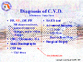
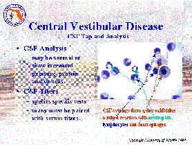 Diagnosis of
central vestibular disease involves the minimal data based for inner ear
disease, but must be expanded to include a chemistry profile, a CSF tap
and analysis (including species specific titers) and, often, advanced brain-imaging
techniques, such as MRI examination. Since CSF cytology is important in
assessing central vestibular disease and advanced imaging techniques are
needed, central vestibular disease crosses "the referral line", the point
in assessing disease which may require the interaction or interpretation
of a neurologist.
Diagnosis of
central vestibular disease involves the minimal data based for inner ear
disease, but must be expanded to include a chemistry profile, a CSF tap
and analysis (including species specific titers) and, often, advanced brain-imaging
techniques, such as MRI examination. Since CSF cytology is important in
assessing central vestibular disease and advanced imaging techniques are
needed, central vestibular disease crosses "the referral line", the point
in assessing disease which may require the interaction or interpretation
of a neurologist.
The treatment and prognosis for central vestibular disease depends upon
the cause. In neoplasia, biopsy may help determine whether radioablative
surgery might be useful. Unfortunately, the brainstem is not an area amenable
to conventional neurosurgery. In small animals, bacterial infections causing
central vestibular disease is uncommon. Rickettsial infection is also rare.
In cats, cryptococcosis may respond to therapy whether with remission or
control of the neurologic signs. In dogs, fungal diseases usually progress
in spite of vigorous treatment. Toxoplasmosis may be controllable for a
period in the dog and treatable in the cat. Canine distemper virus infection
may run its course and stop or be chronic and progressive. FeLV and FIP
infections are generally, rapidly progressive. Granulomatous meningoencephalitis
(GME) will respond temporarily to corticosteroid therapy, but ultimately
progress. Steroid-responsive meningoencephalitis can be controlled with
medication for long periods in the dog. Finally, organophosphate intoxication
and thiamine deficiency 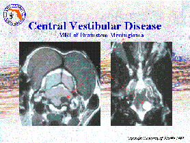 may
respond to appropriate therapy.
may
respond to appropriate therapy.
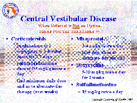 So, while central
vestibular disease has many causes, in the absence of specific disease
processes, there is limited hope for successful treatment and that hope
is often based upon the response of the animal to medical management. The
medical management is largely based upon the responsiveness of the disease
process to corticosteroids. In other words, there are many causes of central
vestibular disease, but often only one treatment approach. If the client
understands this, it is possible to treat central vestibular disease without
a specific diagnosis, realizing that the response to therapy can suggest
whether the disease was "good" or "bad". The treatment approach that I
use when a specific diagnosis cannot be made (either because the patient
is too ill to undergo the diagnostic test or the client cannot afford them)
is to treat with corticosteroids (usually oral prednisolone) and antibiotics
(doxycycline and sulfadimethozine). The prednisolone will reduce inflammation
while the doxycycline can help control bacterial and rickettsial disease
while the sulfadimethozine may help control protozoal infection. I take
a more pro-active approach in cats, since toxoplasmosis and cryptococcosis
might be treatable. Therefore, I prefer to always perform CSF tap and analysis
in cats with central vestibular disease, particularly when they also exhibit
active chorioretinitis. I also add parenteral thiamine therapy when treating
cats.
So, while central
vestibular disease has many causes, in the absence of specific disease
processes, there is limited hope for successful treatment and that hope
is often based upon the response of the animal to medical management. The
medical management is largely based upon the responsiveness of the disease
process to corticosteroids. In other words, there are many causes of central
vestibular disease, but often only one treatment approach. If the client
understands this, it is possible to treat central vestibular disease without
a specific diagnosis, realizing that the response to therapy can suggest
whether the disease was "good" or "bad". The treatment approach that I
use when a specific diagnosis cannot be made (either because the patient
is too ill to undergo the diagnostic test or the client cannot afford them)
is to treat with corticosteroids (usually oral prednisolone) and antibiotics
(doxycycline and sulfadimethozine). The prednisolone will reduce inflammation
while the doxycycline can help control bacterial and rickettsial disease
while the sulfadimethozine may help control protozoal infection. I take
a more pro-active approach in cats, since toxoplasmosis and cryptococcosis
might be treatable. Therefore, I prefer to always perform CSF tap and analysis
in cats with central vestibular disease, particularly when they also exhibit
active chorioretinitis. I also add parenteral thiamine therapy when treating
cats.
We have made important strides in understanding the breadth of central
vestibular diseases. There are new promising approaches which may help
treat more of the diseases than we have previously been able to treat.
New antifungal drugs offer hope in controlling CNS fungal infections. More
potent cytotoxic drugs may help diminish the effects of GME in the dog.
Computer-Assisted Radioablative surgery offers hope in treating brainstem
tumors. On the other hand, we have a long way to go. These new methods
are expensive and not always available to every pet owner. We are, however,
investigating whether natural medicine might be useful adjunctive therapies
in many vestibular diseases. These approaches might reduce the cost and
improve the outcome and prognosis for many patients.
Copyright Dog2Doc.com 1997
All Rights Reserved
 Hop Back to the
Dog2Doc Home Page!!!
Hop Back to the
Dog2Doc Home Page!!!
View
PowerPoint Slide Show on Vestibular Disease
Last updated 27 August 2002