QUADRIPARESIS and QUADRIPLEGIA
by R.M. Clemmons, DVM, PhD
Associate Professor of Neurology & Neurosurgery
Introduction:
Quadriparesis (weakness and ataxia of all 4 limbs) and quadriplegia (paralysis
of all 4 limbs) are common problems in all animals. Once the neurologist,
faced with an animal who has neurologic disease affecting all 4 limbs,
has determined that the lesion is below the foramen magnum (meaning a spinal
cord or peripheral disease), there are 4 possible anatomic locations for
the disease process: 1) if there is UMN dysfunction in all 4 legs, the
lesion is most likely to be in the spinal cord between C1-C5; 2) if there
is LMN dysfunction in the fore legs and UMN dysfunction to the rear legs,
the lesion is severe and involves spinal cord segments C6-T2; 3) if there
is UMN dysfunction to the rear legs and "root signature" (lameness due
to nerve root involvement) in the forelegs, the lesion is mild and affecting
spinal cord segments C6-T2; or, 4) if there is LMN dysfunction in all 4
limbs, the lesion is due to a diffuse LMN disease.
In developing the differential diagnosis for quadriparesis, the basic
mechanisms of disease must be considered along with the signalment and
history. Congenital diseases are not uncommon in the cervical spinal column
of dogs. These include agenesis of the dens (with resultant atlantoaxial
subluxation), blocked vertebra, multiple cartilaginous exostoses, leukoencephalomyelopathy
of Rottweilers, and hereditary ataxia of Jack Russell and Smooth-haired
Fox terriers. In older animals, degenerative intervertebral disc (IVD)
disease, inflammatory meningomyelitis and neoplasia are not uncommon. If
the signs are symmetrical, then nutritional, metabolic and toxic diseases
must be considered. On the other hand, most asymmetrical diseases can be
separated into their most likely causes, which must be included in the
differential. These causes are discospondylitis, meningomyelitis, IVD disease
and neoplasia.
Diagnostic Approach:
Like the rest of the nervous system, the neurologic examination is the
single most important diagnostic method to localize diseases of the cervical
spine, providing an indication from which to make a tentative differential
diagnostic list. On the other hand, localizing diseases in the cervical
spinal column to a specific spinal segment can be difficult, since tests
like the panniculus response cannot be performed there. Hyperpathia can
be difficult to elicit and hyperesthesia is not easily mapped.
The ancillary diagnostic tests for spinal cord disease are similar regardless
of the cause and include the minimum data base, spinal radiographs, EMG,
CSF tap and analysis, myelography and MRI. The minimum data base will often
be normal or may need to be expanded based upon the physical and neurologic
examinations. In older patients, routine chest and abdominal radiographs
and abdominal ultrasound may help make a diagnosis of the cervical disease
or assist in making the prognosis. Spinal radiographs may show signs of
degenerative disc disease, congenital malformation, spinal arthritis or
discospondylitis. The later disease being the only disease diagnosis which
can be made on plain spinal radiographs. The other diseases will need additional
imaging techniques to confirm that they are the source of the problem.
In acute diseases, the EMG may not help identify denervation until 5-7
days have past; however, nerve conduction velocity studies may help identify
damaged nerves or diffuse LMN disorders. On the other hand, in chronic
diseases, the EMG may help to localize the disease process, so that radiographs
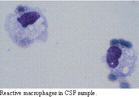
can concentrate on the lesion. The CSF tap can help determine the presence
of inflammation or infection in cervical diseases. The problem of inflammatory
myelitis is increasing, making CSF tap and analysis critical in assessing
cervical neurologic disease. Even when other neurologic conditions are
identified, myelitis may be present. Unfortunately, many patients are treated
with corticosteroids before being adequately worked-up for cervical disease.
The work-up performed in the face of the steroids may be erroneous. As
such, surgical intervention may be performed, only later to discover the
cause of neck pain was inflammatory meningomyelitis. Spinal myelography
helps to contrast the spinal cord when looking for mass lesions. It can
be an extremely valuable diagnostic aid in determining the need for surgical
intervention and what surgical approach is best. In cervical vertebral
malformation complex, the lesion is dynamic. The only imaging technique
which can provide dynamic views is the myelogram. Myelography, therefore,
remains the single most important imaging technique for assessing surgical
diseases in the cervical spine. When the myelographic data is lacking or
when it is not clear what the lesion represents, MRI can add diagnostic
detail. MRI may be important in assessing neoplastic disease processes,
including nerve root tumors. The sequence of diagnostic tests logically
follows the pattern of minimum data base, EMG, spinal radiographs, CSF
tap, myelography and, finally, MRI. If an accurate diagnosis is made along
the way, the remaining test may not be needed.
Specific Disorders
Meningomyelitis:
As stated before, meningomyelitis appears to be on the rise. Twenty years
ago, it was rare to diagnose meningomyelitis and most of these were secondary
to canine distemper virus with the remainder being due to toxoplasmosis.
Today, it is almost impossible to deal with animals with neck pain and
not be suspicious of meningomyelitis. For this reason, even with signs
of early degenerative disc disease, I do not consider surgery until I have
ruled-out meningitis. While some neurologists are unconcerned about performing
myelography on patients who have meningomyelitis, most contrast agents
are inflammatory by nature. In the face on meningomyelitis, myelography
can exacerbate the clinical signs and is, therefore, generally contraindicated
in meningomyelitis.
 The clinical
signs of meningomyelitis are, generally, neck pain and asymmetrical neurologic
deficits. The deficits depend upon which pathways are involved in the disease
process. The signs are usually progressive, but may develop acutely. In
dogs and cats, the causes of meningomyelitis are, in order of likelihood,
viral, inflammatory, protozoal, fungal, rickettsial and bacterial diseases.
The viral disease most commonly seen in dogs is canine distemper (even
in vaccinated dogs). In cats, feline leukemia virus (FeLV), feline infectious
peritonitis (FIP) and feline immunodeficiency virus (FIV) are the most
common viral infections. Toxoplasmosis can occur in both dogs and cats,
while dog also may develop Neospora caninum infections. Aspergillosis
is not uncommon in dogs, while cryptococcosis is more common in cats. Cats
do not appear to have rickettsial diseases, but dogs have been shown to
develop meningomyelitis from both ehrlichiosis and Rocky Mountain spotted
fever. Titers for these agents should be performed on the serum and/or
CSF when presented with meningomyelitis.
The clinical
signs of meningomyelitis are, generally, neck pain and asymmetrical neurologic
deficits. The deficits depend upon which pathways are involved in the disease
process. The signs are usually progressive, but may develop acutely. In
dogs and cats, the causes of meningomyelitis are, in order of likelihood,
viral, inflammatory, protozoal, fungal, rickettsial and bacterial diseases.
The viral disease most commonly seen in dogs is canine distemper (even
in vaccinated dogs). In cats, feline leukemia virus (FeLV), feline infectious
peritonitis (FIP) and feline immunodeficiency virus (FIV) are the most
common viral infections. Toxoplasmosis can occur in both dogs and cats,
while dog also may develop Neospora caninum infections. Aspergillosis
is not uncommon in dogs, while cryptococcosis is more common in cats. Cats
do not appear to have rickettsial diseases, but dogs have been shown to
develop meningomyelitis from both ehrlichiosis and Rocky Mountain spotted
fever. Titers for these agents should be performed on the serum and/or
CSF when presented with meningomyelitis.
The diagnosis is made on CSF tap and analysis. Generally, we approach
animals with neck pain and quadriparesis by performing a minimum data base
including a CBC, chemistry profile, urinalysis and appropriate radiographs.
With the CBC, we run plasma fibrinogen levels. This is a crude estimate
of systemic inflammation, but a valuable tool in assessing the potential
for meningomyelitis. It may be the only abnormality noted in the CBC. Once
the minimal data base is evaluated, we proceed with anesthesia and CSF
tap. While this is being processed, spinal radiographs are taken. If the
CSF indicates inflammation by increase in cells and protein and the survey
radiographs do not demonstrate significant findings, we then treat the
inflammation rather than proceed with myelography. Based upon the response
to therapy, we reassess the need for further tests. CSF titers are submitted
for the relevant infectious agents providing confirmation of the specific
disease causing organism. In those cases where a specific disease causing
organism can be found, the treatment is adjusted appropriately. When no
organism is found, the tentative diagnosis of inflammatory meningomyelitis
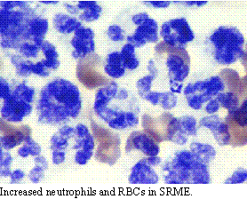
is made. Many newer forms of meningomyelitis are now recognized including
steroid-responsive meningomyelitis. This is usually associated with an
increase in blood vessel fragility and may lead to an apparently blood-contaminated
CSF tap. On examination, however, there is a marked increase in non-degenerative
neutrophils in the CSF.
 As in beagles
with necrotizing vasculitis (beagle neck pain syndrome), many patients
with steroid-responsive meningomyelitis have elevations in alpha 2 globulins
on serum electrophoresis. Steroid-responsive meningomyelitis probably represents
a form of vasculitis which results in inflammation in the CNS. Conventional
therapy with corticosteroids will not always resolve this condition, since
steroids only suppress the symptoms of the disease. Although some dogs
recover from this disease following corticosteroid management, many would
probably benefit from alternative therapy. Conventional therapy involves
giving prednisolone at 1 mg/kg/day in three divided doses. Once the signs
resolve (usually within 72 hours), the dosage is reduced to twice a day.
This is further reduced to daily medication in the morning and, finally,
to alternate day therapy. We find that many patients will benefit from
anti-oxidant therapy, including vitamin E, vitamin C and selenium. Additional
medications of benefit include omega-3-fatty acids, ginkgo biloba extract
and green tea. When pain is present, garlic, ginger and feverfew may help
reduce the inflammation without causing additional gastrointestinal signs.
Some patients will be relieved by the alternative medication, reducing
or replacing the corticosteroid.
As in beagles
with necrotizing vasculitis (beagle neck pain syndrome), many patients
with steroid-responsive meningomyelitis have elevations in alpha 2 globulins
on serum electrophoresis. Steroid-responsive meningomyelitis probably represents
a form of vasculitis which results in inflammation in the CNS. Conventional
therapy with corticosteroids will not always resolve this condition, since
steroids only suppress the symptoms of the disease. Although some dogs
recover from this disease following corticosteroid management, many would
probably benefit from alternative therapy. Conventional therapy involves
giving prednisolone at 1 mg/kg/day in three divided doses. Once the signs
resolve (usually within 72 hours), the dosage is reduced to twice a day.
This is further reduced to daily medication in the morning and, finally,
to alternate day therapy. We find that many patients will benefit from
anti-oxidant therapy, including vitamin E, vitamin C and selenium. Additional
medications of benefit include omega-3-fatty acids, ginkgo biloba extract
and green tea. When pain is present, garlic, ginger and feverfew may help
reduce the inflammation without causing additional gastrointestinal signs.
Some patients will be relieved by the alternative medication, reducing
or replacing the corticosteroid.
Discospondylitis:
Discospondylitis represents an infection of the vertebrae associated with
abscessation of the intervertebral space. It may be secondary to a migrating
foreign body; but, often, no specific source of the infections is found.
It is thought that, in most cases, there is a hematogenous spread of the
infection which isolates into a degenerative disc. Although some cases
are associated with vegetative endocarditis, most do not demonstrate a
source of infection. It may be that agents enter through inflamed tissues
associated with periodontal disease. In cases where there is persistent
or intermittent fever, blood cultures may provide information about the
infection. This is, however, less common than finding the organism in the
urine.
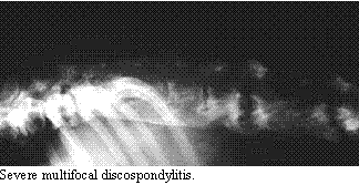
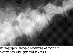 The primary complaint
in discospondylitis is pain at the site of infection. In severe cases,
quadriparesis and anorexia may be present with cervical discospondylitis.
The diagnosis is confirmed by routine spinal radiographs showing characteristic
lysis and sclerosis of the adjacent endplates of the vertebrae. This is
one of the few neurologic conditions where the diagnosis can be made on
routine radiographic examination. The minimum data base includes a CBC
(with a marker of inflammation such as the plasma fibrinogen level), urinalysis
(with culture), fecal examination, Brucella canis titer, and spinal
radiographs. Chest radiographs and echocardiography may be indicated if
there is a heart murmur. Since the radiography changes may not occur until
2-3 weeks from the start of clinical signs, repeat radiographic examination
is indicated when discospondylitis is high on the differential list. The
CBC may reflect changes consistent with infection (including neutrophilia)
or be normal. On of the important monitors is the marker of inflammation.
We use fibrinogen, since it is easy and inexpensive to run. When the fibrinogen
levels are elevated, this is a good indicator of a disease with much tissue
reaction. On the other hand, when the fibrinogen is low, I am particularly
concerned about the possibility of fungal disease. In the later case, I
usually perform a routine chest radiograph looking for discospondylitic-like
lesions between the sternebrae. When lesions are also present between the
sternebrae, most often fungal infection is the cause of the discospondylitis
lesions.
The primary complaint
in discospondylitis is pain at the site of infection. In severe cases,
quadriparesis and anorexia may be present with cervical discospondylitis.
The diagnosis is confirmed by routine spinal radiographs showing characteristic
lysis and sclerosis of the adjacent endplates of the vertebrae. This is
one of the few neurologic conditions where the diagnosis can be made on
routine radiographic examination. The minimum data base includes a CBC
(with a marker of inflammation such as the plasma fibrinogen level), urinalysis
(with culture), fecal examination, Brucella canis titer, and spinal
radiographs. Chest radiographs and echocardiography may be indicated if
there is a heart murmur. Since the radiography changes may not occur until
2-3 weeks from the start of clinical signs, repeat radiographic examination
is indicated when discospondylitis is high on the differential list. The
CBC may reflect changes consistent with infection (including neutrophilia)
or be normal. On of the important monitors is the marker of inflammation.
We use fibrinogen, since it is easy and inexpensive to run. When the fibrinogen
levels are elevated, this is a good indicator of a disease with much tissue
reaction. On the other hand, when the fibrinogen is low, I am particularly
concerned about the possibility of fungal disease. In the later case, I
usually perform a routine chest radiograph looking for discospondylitic-like
lesions between the sternebrae. When lesions are also present between the
sternebrae, most often fungal infection is the cause of the discospondylitis
lesions.
 The causative
agents are bacteria (Staphylococcus, Streptococcus and Corynebacterium
are the most common, although Brucella can occasionally be seen
as a cause), parasitic (Spirocerca lupi in thoracic discospondylitis),
and fungal (Aspergillus and Nocardia). As such, the treatment
and prognosis vary depending upon the organism causing the infection. Parasitic
infections are rare except in the Southwestern US and usually represent
advanced cases of parasitism. Brucella canis infection is not uncommon,
but much less so than the other bacterial causes. When Brucella
appears to be the cause, antibiotic therapy must take this into account
(usually, I use doxycycline). Fungal infections with Aspergillus
do not respond well to antifungal drugs. Recently, there have been reports
of controlling the infection for extended period using itraconazole. I
use raw garlic in hopes that it will help control the problem.
The causative
agents are bacteria (Staphylococcus, Streptococcus and Corynebacterium
are the most common, although Brucella can occasionally be seen
as a cause), parasitic (Spirocerca lupi in thoracic discospondylitis),
and fungal (Aspergillus and Nocardia). As such, the treatment
and prognosis vary depending upon the organism causing the infection. Parasitic
infections are rare except in the Southwestern US and usually represent
advanced cases of parasitism. Brucella canis infection is not uncommon,
but much less so than the other bacterial causes. When Brucella
appears to be the cause, antibiotic therapy must take this into account
(usually, I use doxycycline). Fungal infections with Aspergillus
do not respond well to antifungal drugs. Recently, there have been reports
of controlling the infection for extended period using itraconazole. I
use raw garlic in hopes that it will help control the problem.
By in large, the most common causes of discospondylitis are secondary
to bacteria which can be treated using a combination of sulfa drugs (sulfadimethozine,
15 mg/kg every 12 hours) and either cephalosporins (22 mg/kg every 8-12
hours) or enrofloxacin (5-7.5 mg/kg every 12 hours). I prefer the former
combination and treat the infection for a minimum of 6-8 weeks. Radiographic
repair usually lags behind remission of the infection; however, following
the response to therapy and continuing therapy beyond the time of radiographic
quiescence seem the best policy. In cases which do not respond, the urine
should be reexamined and abdominal ultrasound of the kidneys performed,
looking for evidence that fungal disease was the real cause. Rarely, the
infection will result in bony compression or instability requiring surgical
intervention. Most often, spinal cord compression is the result of soft
tissue inflammation which subsides quickly with appropriate antibiotic
therapy.
Cervical Vertebral Malformation Complex:
 Wobbler's disease
occurs in young and old animals. In young animals, it appears to be secondary
to inherited malformation and mis-articulation of the cervical vertebrae
which is accentuated by high protein diets. In older animals, it appears
to be secondary to chronic degenerative disc disease. Although other large
breeds can be affected, it is said to be a disease of young Great Danes
and old Doberman Pinchers. When a Doberman Pincher presents with signs
of rear leg ataxia with "root signature" in the forelegs, there is a high
probability that the dog has Wobbler's disease.
Wobbler's disease
occurs in young and old animals. In young animals, it appears to be secondary
to inherited malformation and mis-articulation of the cervical vertebrae
which is accentuated by high protein diets. In older animals, it appears
to be secondary to chronic degenerative disc disease. Although other large
breeds can be affected, it is said to be a disease of young Great Danes
and old Doberman Pinchers. When a Doberman Pincher presents with signs
of rear leg ataxia with "root signature" in the forelegs, there is a high
probability that the dog has Wobbler's disease.
The onset of clinical signs can be acute or slow and insidious. There
is evidence of ataxia in all four limbs with the pelvic limbs being more
affected. There will be both conscious and unconscious proprioceptive dysfunction
with a wide-based stance in the rear legs. The forelegs may show a stiff
and stilted gait with atrophy or fasciculations of the deltoideus, biceps
and infra- and supraspinatus muscles. There is usually some degree of neck
pain on palpation and neck manipulations. One sign of this is a reluctance
to hop medially with the forelegs.
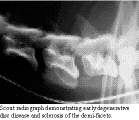
 The diagnosis
can be suspicioned on survey radiographs of the neck, looking for narrowed
IVD spaces and sclerosis of the demi-facets. CSF analysis is usually within
normal limits, although a small number of cases will show a mild increase
in cells (4-10 cells/µl) and protein (25-35 mg/ml). EMG can help
confirm the location and the denervation of the muscles with fasciculations.
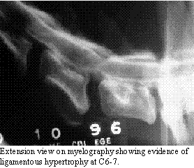
The diagnosis in confirmed on myelography, which shows evidence of IVD
protrusion and the presence of ligamentous or bony intrusion into the neural
canal. Since CVM represents a dynamic lesion, myelography with mildly flexed
and extended views is the diagnostic technique of choice. It is also important
to take a "lazy" lateral view, since stretching the neck can reduce the
lesion so as to overlook it. If the lesion is alleviated with flexion and
accentuated on extension, the problem is partially due to ligamentous hypertrophy.
On the other hand, if flexion and extension do not affect the lesion, it
is probably secondary to IVD protrusion. I feel that a single lesion is
better than multiple ones. Further, an IVD protrusion is less problematic
than one with ligamentous hypertrophy.
The diagnosis
can be suspicioned on survey radiographs of the neck, looking for narrowed
IVD spaces and sclerosis of the demi-facets. CSF analysis is usually within
normal limits, although a small number of cases will show a mild increase
in cells (4-10 cells/µl) and protein (25-35 mg/ml). EMG can help
confirm the location and the denervation of the muscles with fasciculations.
The diagnosis in confirmed on myelography, which shows evidence of IVD
protrusion and the presence of ligamentous or bony intrusion into the neural
canal. Since CVM represents a dynamic lesion, myelography with mildly flexed
and extended views is the diagnostic technique of choice. It is also important
to take a "lazy" lateral view, since stretching the neck can reduce the
lesion so as to overlook it. If the lesion is alleviated with flexion and
accentuated on extension, the problem is partially due to ligamentous hypertrophy.
On the other hand, if flexion and extension do not affect the lesion, it
is probably secondary to IVD protrusion. I feel that a single lesion is
better than multiple ones. Further, an IVD protrusion is less problematic
than one with ligamentous hypertrophy.
 The treatment of CVM is surgery. In cases where surgery is not possible
(patient has complications or is elderly), medical management with prednisolone
and diazepam may provide temporary relief. However, in the absence of compelling
reasons not to perform surgery, surgical decompression is needed. There
are several surgical techniques available to treat Wobbler's disease including
dorsal laminectomy, ventral slot and ventral slot with distraction (by
various means). In cases of multiple lesions, dorsal laminectomy was the
method of choice, in the past. Dorsal laminectomy has risks and the success
rate is the lowest of methods for correcting CVM. In qualified hands, it
is still a good technique. The overall success is around 75% with a 20-25%
morbidity and a 5-10% mortality. Large breeds do not tolerate dorsal laminectomy
well. Ventral slot is excellent for IVD protrusion, but increases compression
from ligamentous hypertrophy. In simple IVD protrusion, ventral slot has
a 90-95% success rate with a 5% morbidity and <1% mortality. The morbidity
and mortality increase for ventral slots when ligamentous hypertrophy is
present. When ligamentous hypertrophy is present, ventral slot alone is
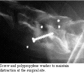
generally inadequate to correct the problem. A number of techniques have
been described to perform a ventral slot and maintain distraction across
the IVD space. These methods include various implants from Harrington rods
to screws (or pins) and methylmethacrylate. The method we use is a modified
"screw and washer" technique. The washer we use is a polypropylene ring
made from the end of an endotracheal tube. This is packed with bone graft
and the screw stabilizes the implant while fusion takes place. In this
method, the "slot" is performed by discectomy without removing the endplates.
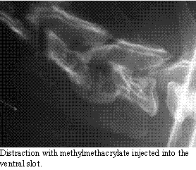
It is my belief that fusion is the goal of CVM surgery and this is best
done in distraction. Another popular method (which I do not believe provides
adequate fusion) is to inject methylmethacrylate between the vertebrae
at the ventral slot site. This can be used as a salvage procedure.
The treatment of CVM is surgery. In cases where surgery is not possible
(patient has complications or is elderly), medical management with prednisolone
and diazepam may provide temporary relief. However, in the absence of compelling
reasons not to perform surgery, surgical decompression is needed. There
are several surgical techniques available to treat Wobbler's disease including
dorsal laminectomy, ventral slot and ventral slot with distraction (by
various means). In cases of multiple lesions, dorsal laminectomy was the
method of choice, in the past. Dorsal laminectomy has risks and the success
rate is the lowest of methods for correcting CVM. In qualified hands, it
is still a good technique. The overall success is around 75% with a 20-25%
morbidity and a 5-10% mortality. Large breeds do not tolerate dorsal laminectomy
well. Ventral slot is excellent for IVD protrusion, but increases compression
from ligamentous hypertrophy. In simple IVD protrusion, ventral slot has
a 90-95% success rate with a 5% morbidity and <1% mortality. The morbidity
and mortality increase for ventral slots when ligamentous hypertrophy is
present. When ligamentous hypertrophy is present, ventral slot alone is
generally inadequate to correct the problem. A number of techniques have
been described to perform a ventral slot and maintain distraction across
the IVD space. These methods include various implants from Harrington rods
to screws (or pins) and methylmethacrylate. The method we use is a modified
"screw and washer" technique. The washer we use is a polypropylene ring
made from the end of an endotracheal tube. This is packed with bone graft
and the screw stabilizes the implant while fusion takes place. In this
method, the "slot" is performed by discectomy without removing the endplates.
It is my belief that fusion is the goal of CVM surgery and this is best
done in distraction. Another popular method (which I do not believe provides
adequate fusion) is to inject methylmethacrylate between the vertebrae
at the ventral slot site. This can be used as a salvage procedure.
Following surgery, the patient should be kept quite for 30 days and
supported with a neck brace and bandage. After the first month, the activity
level is gradually returned to normal. Depending upon the severity of the
initial damage, most patients will improve, reaching 80% of their recovery
in the first 3 months. There is a potential for the "domino" effect, whereby
the IVD on 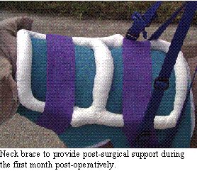 either
side of the surgery site will develop problems in 6 months to 2 years following
the initial correction. I find that this is more often when the beginnings
of CVM were present in the beginning. We now are much more aggressive than
in the past, fixing multiple lesions from the start.
either
side of the surgery site will develop problems in 6 months to 2 years following
the initial correction. I find that this is more often when the beginnings
of CVM were present in the beginning. We now are much more aggressive than
in the past, fixing multiple lesions from the start.
Copyright Dog2Doc.com 1997
All Rights Reserved
 Hop back to the Dog2Doc Home Page!
Last updated 27 August 2002